Mammograms are currently the industry standard test for breast cancer. A mammogram is an x-ray that allows a specialist to examine the breast tissue for any suspicious areas. The breast is exposed to a dose of iodizing radiation that produces an image of the breast tissue.

There are two issues with this. First, you are exposing your body to radiation needlessly. Exposure to radiation over time has been shown to cause DNA level cellular mutations (cancer). Secondly by applying that much pressure, if there is a tumor (which is an encapsulation of cells, possibly cancerous (mutated/toxic) your body has worked hard to cordon those of, protecting the other cells from them. The pressure exerted by a mammogram machine (up to 50lbs. per breast) is enough that it could burst that encasement, spreading the cells, even possibly down into the lymph nodes causing further problems that may not otherwise have ever existed.
A more recent development in technology which is far less invasive and there for safer, is to use Thermographic Imaging.
How does Thermographic Imaging (Thermography) work?
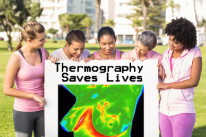
Thermography is a form of infrared imaging using cameras that “read” the entire infrared range of the electromagnetic spectrum and produce images. It is a noninvasive test that can be done using the body’s heat map in order to detect abnormalities, including (but not limited to) cancer.
“It is widely acknowledged that cancers, even in their earliest stages, need nutrients to maintain or accelerate their growth. In order to facilitate this process, blood vessels are caused to remain open, inactive blood vessels are activated, and new ones are formed through a process known as neoangiogenesis. This vascular process causes an increase in surface temperature in the affected regions, which can be viewed with infrared imaging cameras. Additionally, the newly formed or activated blood vessels have a distinct appearance, which thermography can detect.” – Dr. Philip Getson, D.O. http://tdinj.com/about-tdi/dr-philip-getson/
He goes on to add that “Since thermal imaging detects changes at the cellular level, studies suggest that this test can detect activity 8 to 10 years before any other test. This makes it unique in that it affords us the opportunity to view changes before the actual formation of the tumor.”
In 2009 the United States Preventative Services Task Force stated in the New Mammography Guidelines that mammograms should begin at 50 rather than the previously stated 40 stating that “Risk of United States Preventative Services Task Force additional and unnecessary testing far outweighed the benefits of annual mammograms”. (http://www.uspreventiveservicestaskforce.org/)
With thermographic imaging, there is no risk. It can be performed on pregnant and breast feeding women as well. This procedure is safe not only for breast tissue, but to use to scan the entire body if needed. Thermography is an incredibly good choice for younger women’s breasts, as they are usually denser. It doesn’t read fibrocystic tissue, breast implants or scars as needing further investigation as sometimes a mammogram can. Additionally it can also detect changes in the cells in the under arm area lymph nodes. This is an area that mammography isn’t always accurate in screening.
Thermography encourages a healthy proactive approach to breast health. Instead of just screening for breast cancer, a thermogram can give you an overall reading of breast tissue health. It has the potential to detect breast cell abnormalities long before mammography can detect cancer, when done properly. This allows you to implement lifestyle and diet changes that can improve the health of your breasts proactively instead of waiting for a cancer diagnosis.
If you are age 40 or over and your doctor is recommending you begin yearly mammograms do a little research of your own into the options before scheduling an appointment.
If you are looking for a General Practitioner, Gynecologist or Oncologist here in Lee County, please visit www.ipalc.org and search our database.
Share on Facebook


 Southwest Florida Medicine.com is dedicated to bringing you the very best health information available today!
Subscribe or check back regularly!